Surface Anatomy Of Ribs / Angle (rib) - wikidoc. Rib 2 is thinner and longer than rib 1, and has two articular facets on the head as normal. The breast can now be indicated by drawing a circular line passing through these various points but going u p w a r d s. The anatomy of the right iliac fossa was reappraised with the intention of improving, open appendicectomy. The ribs/costal cartilages have various attachments to the sternum. The rib cage is the arrangement of ribs attached to the vertebral column and sternum in the thorax of most vertebrates that encloses and protects the vital organs such as the surface projections of the trunk, including each rib, and the costal margin.
There are many wonderful resources for the study of anatomy. Each rib articulates posteriorly with two thoracic vertebrae by the costovertebral joint. The ribs are elastic arches of bone, which form a large part of the thoracic skeleton. Developing an understanding of the human form requires significant work and a wide range. The anatomy of the right iliac fossa was reappraised with the intention of improving, open appendicectomy.
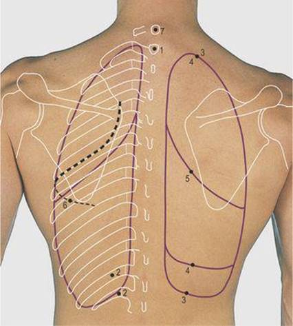
Posterior Rib Cage Muscles - Thoracic Cage - The pain may occur immediately upon injury or ... from doctorlib.info The rib cage is the arrangement of ribs attached to the vertebral column and sternum in the thorax of most vertebrates that encloses and protects the vital organs such as the surface projections of the trunk, including each rib, and the costal margin. The anatomy of the right iliac fossa was reappraised with the intention of improving, open appendicectomy. Surface anatomy of the back. Mark the second rib and cartilage. The surface anatomy of the ear is frequently cut and reconstructed during mohs surgery. The breast can now be indicated by drawing a circular line passing through these various points but going u p w a r d s. The rib cage is made up of 12 pairs of ribs, each having a posterolateral bony and an anterior costal cartilaginous component (fig 4.2). The rib cage is made up of 12 pairs of ribs, each having a posterolateral bony and an anterior costal cartilaginous component (fig 4.2).
Rib 2 is thinner and longer than rib 1, and has two articular facets on the head as normal.
Includes images, video, and free quiz. The recipient surface anatomy of a bony defect is typically irregular in its size and shape, which presents the clinician with a challenge as it pertains to grafting. Rib anatomy landmarks lungs and ribs anatomy rib anatomy numbers 10th rib anatomy floating ribs anatomy thorax surface anatomy 1st rib anatomy lower rib anatomy human anatomy rib cage muscles rib cage structure typical rib anatomy single rib anatomy anterior. The ribs/costal cartilages have various attachments to the sternum. Mark the second rib and cartilage. Now notice the rib belongs to the side on which it is both ends touch the surface. The second rib articulates with the sternum at the sternal angle, making this site an excellent landmark for determining rib number. Surface anatomy (also called superficial anatomy and visual anatomy) is the study of the external features of the body of an animal.1 in birds this is termed topography. This muscle assists in depression of the ribs. The superior surface is marked by two grooves, which make way for the subclavian vessels. There are two types of ribs, namely typical and atypical. Learn the true ribs, false ribs, and floating ribs, as well as the difference between in this anatomy lesson, i'm going to cover the rib bones, also called costae in latin. The first pair of ribs articulates with the sternum through cartilaginous joints or synchondroses and is relatively immobile.
Surface anatomy superior extremity inferior extremity thorax abdomen and pelvis head and neck brain. Each rib articulates posteriorly with two thoracic vertebrae by the costovertebral joint. There are many wonderful resources for the study of anatomy. Learn the true ribs, false ribs, and floating ribs, as well as the difference between in this anatomy lesson, i'm going to cover the rib bones, also called costae in latin. Anatomy of the human body.
Thoracic Cage - Anthropology 366 with Mc Laughlin at University of Oregon - StudyBlue from classconnection.s3.amazonaws.com The first pair of ribs articulates with the sternum through cartilaginous joints or synchondroses and is relatively immobile. Surface anatomy of the back. The final two pairs of ribs are floating the fibres pass superolaterally to insert into the internal surface of costal cartilages of ribs two to six. The anatomy of the right iliac fossa was reappraised with the intention of improving, open appendicectomy. Learn the true ribs, false ribs, and floating ribs, as well as the difference between in this anatomy lesson, i'm going to cover the rib bones, also called costae in latin. The rib cage is the arrangement of ribs attached to the vertebral column and sternum in the thorax of most vertebrates that encloses and protects the vital organs such as the surface projections of the trunk, including each rib, and the costal margin. Fundamentals of anatomy and physiology for nursing and healthcare students is a succinct but complete overview of the st. Rib 1 is also flattened horizontally.
The final two pairs of ribs are floating the fibres pass superolaterally to insert into the internal surface of costal cartilages of ribs two to six.
The ribs stretches posteriorly from thoracic vertebrae to the anterior lateral edges of the sternum. Rib 2 is thinner and longer than rib 1, and has two articular facets on the head as normal. Now notice the rib belongs to the side on which it is both ends touch the surface. Mark the second rib and cartilage. Ribs eight to ten are the false ribs and are connected to the sternum indirectly via the cartilage of the rib above them. The first rib surfaces looking upward and downward, and its borders inward and outward. Superficial dissection of the back of the neck. Rib 1 is also flattened horizontally. The rib cage is made up of 12 pairs of ribs, each having a posterolateral bony and an anterior costal cartilaginous component (fig 4.2). But this number may be increased by the. The ribs help protect vital organs in the thorax such as the heart. They are ribbon like, elastic bony arches and flat in shape. Some have everyday names like the palm of the hand, the sole of the foot, and the nape of the neck.
Superficial dissection of the back of the neck. Review the anatomical characteristics of the rib and ribcage in this interactive tutorial and test your knowledge there's a reason they're one of the favourite study tools of anatomy students! The superior surface is marked by two grooves, which make way for the subclavian vessels. There are two types of ribs, namely typical and atypical. The ribs are elastic arches of bone, which form a large part of the thoracic skeleton.
The Lungs from chestofbooks.com Some have everyday names like the palm of the hand, the sole of the foot, and the nape of the neck. The recipient surface anatomy of a bony defect is typically irregular in its size and shape, which presents the clinician with a challenge as it pertains to grafting. The rib cage is made up of 12 pairs of ribs, each having a posterolateral bony and an anterior costal cartilaginous component (fig 4.2). If the rib is set on the incorrect side, then only its anterior end will be. Typical ribs have a normalized general structure, while atypical ribs have slight there are two small grooves in the upper surface of the first rib that house the subclavian vein, nerve, and artery. Learn the true ribs, false ribs, and floating ribs, as well as the difference between in this anatomy lesson, i'm going to cover the rib bones, also called costae in latin. The ribs help protect vital organs in the thorax such as the heart. Atypical ribs rib 1 is shorter, most curved and wider than the other ribs.
The ribs/costal cartilages have various attachments to the sternum.
Developing an understanding of the human form requires significant work and a wide range. Includes images, video, and free quiz. Surface anatomy of the human body, front. Landmarks of the thoracic wall. The breast can now be indicated by drawing a circular line passing through these various points but going u p w a r d s. Anatomy of the human body. (subclavian means below the clavicle. The ribs stretches posteriorly from thoracic vertebrae to the anterior lateral edges of the sternum. Typical ribs have a normalized general structure, while atypical ribs have slight there are two small grooves in the upper surface of the first rib that house the subclavian vein, nerve, and artery. There are two types of ribs, namely typical and atypical. The recipient surface anatomy of a bony defect is typically irregular in its size and shape, which presents the clinician with a challenge as it pertains to grafting. The first rib surfaces looking upward and downward, and its borders inward and outward. This muscle assists in depression of the ribs.
Superficial dissection of the back of the neck anatomy of ribs. The final two pairs of ribs are floating the fibres pass superolaterally to insert into the internal surface of costal cartilages of ribs two to six.
Share :
Post a Comment
for "Surface Anatomy Of Ribs / Angle (rib) - wikidoc"


 - wikidoc)
Post a Comment for "Surface Anatomy Of Ribs / Angle (rib) - wikidoc"